العودة إلى الصفحة الرئيسية
820К+ مريضًا حصلوا على المساعدة منذ عام 2014
50 دول
1,500 عيادات
6K+ تقييمات
3K+ طبيب مؤهل

ما هي تكلفة تضيق القحف إجراءات التشخيص والعلاج في الهند؟ اعرف الآن

السعر يقدم بعد الطلب

مزاياك وضماناتك مع Bookimed

أسعار مباشرة

Bookimed لا يضيف رسوماً إضافية على أسعار علاج تضيق القحف. الأسعار مأخوذة من قوائم الأسعار الرسمية للعيادات. تدفع مباشرة في العيادة مقابل علاجك عند وصولك إلى البلد.

عيادات وأطباء معتمدون فقط

Bookimed ملتزم بسلامتك. نحن نعمل فقط مع المؤسسات الطبية التي تحافظ على معايير دولية عالية في علاج تضيق القحف ولديها التراخيص اللازمة لخدمة المرضى الدوليين في جميع أنحاء العالم.

مساعدة مجانية على مدار الساعة

Bookimed يقدم مساعدة خبراء مجانية. منسق طبي شخصي يدعمك قبل وأثناء وبعد العلاج، ويحل أي مشاكل. لن تكون وحيداً أبداً في رحلة علاج تضيق القحف.

لماذا نحن؟

منسقك الطبي الشخصي في Bookimed

  • يدعمك في كل مرحلة
  • المساعدة في اختيار العيادة الطبية المناسبة والطبيب المناسب
  • يضمن الوصول السريع والمريح إلى المعلومات

اكتشف أفضل تضيق القحف العيادات في الهند: 21 خيارات معتمدة والأسعار

تم تصنيف العيادات بواسطة نظام Bookimed الذكي، باستخدام تحليل علوم البيانات عبر 5 معايير رئيسية.
Manipal Hospitals
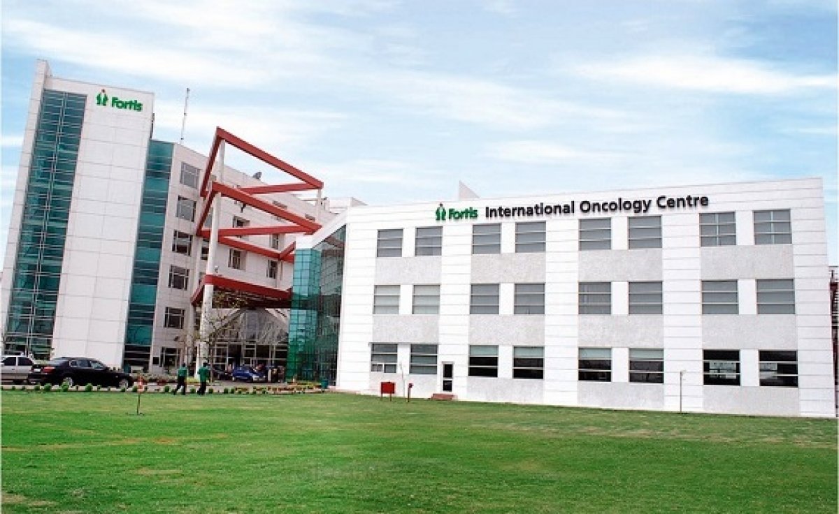
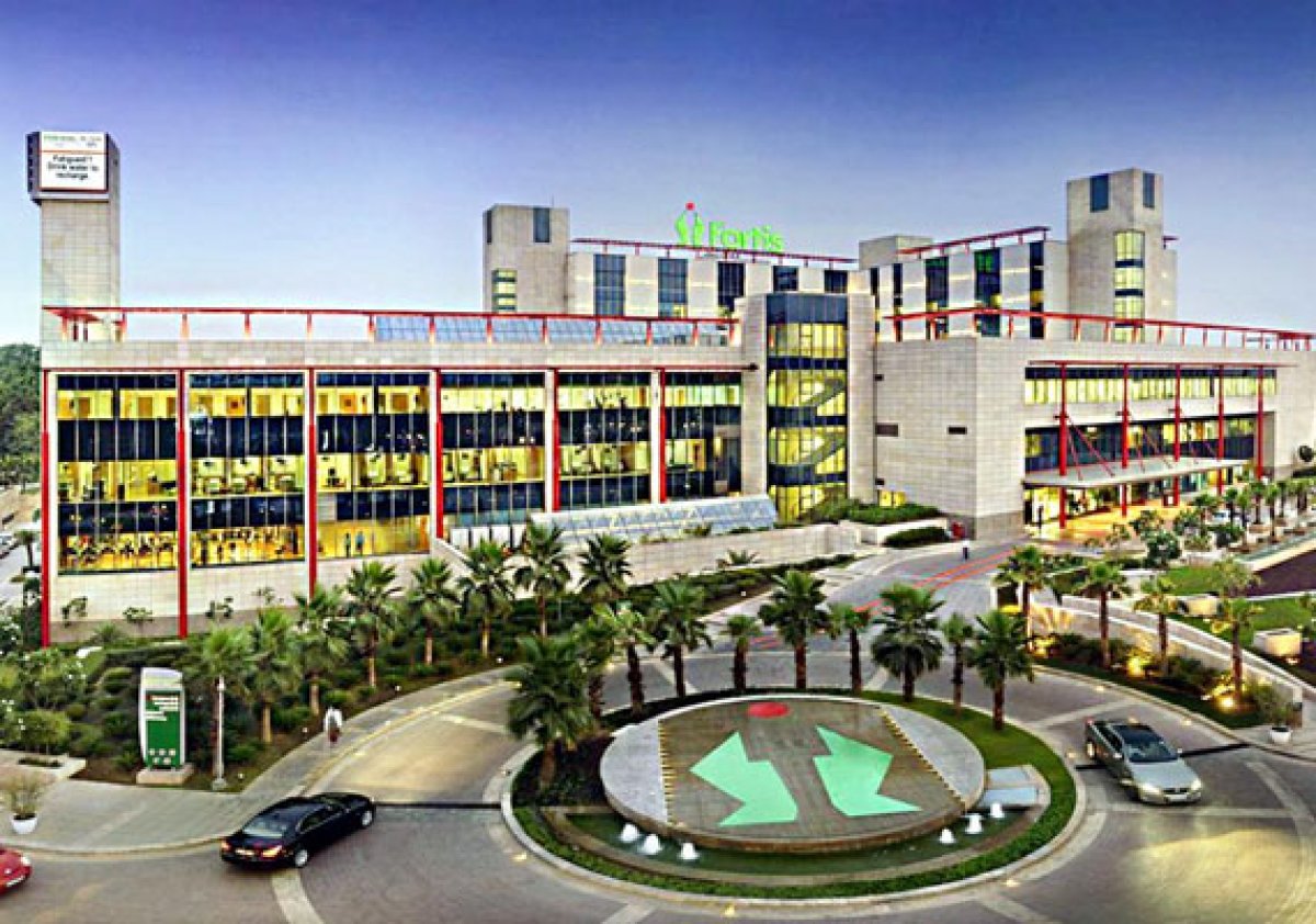
Fortis Gurgaon
Global Hospital Chennai
Aster CMI Hospital
لقد شاهدتم 5 من 21 عيادات

مشاركة المحتوى

Twitter XLinkedinFacebookWhatsAppEmail
أحصل على الرابط

قصص فيديو من مرضى Bookimed

Dayana
جمعت بين إجازتي في أنطاليا والفحص الطبي.
إجراء: فحص صحة الإناث (انظر فحص النساء)
Igor
كان رائعاً! التنقلات والإقامة والعلاج - كل شيء مشمول.
إجراء: زرع الأسنان
مستشفى: WestDent Clinic
Marina
قام Bookimed بكل شيء من أجلي. لم أضطر للقلق بشأن أي شيء.
إجراء: فحص صحة الإناث (انظر فحص النساء)
مستشفى: Severance Hospital
محدث: 05/27/2022
تأليف
Anna Leonova
Anna Leonova
رئيس فريق تسويق المحتوى
محررة طبية معتمدة لديها خبرة تزيد عن 10 سنوات، طوّرت محتوى Bookimed الموثوق، مدعومة بماجستير في علم اللغة ومقابلات مع خبراء طبيين من جميع أنحاء العالم.
تمت المراجعة من قبل المستشار الطبي في Bookimed
فهد مولود
محرر طبي وعالم بيانات
طبيب عام. حاصل على 4 جوائز علمية. خدم في غرب آسيا. رئيس الفريق الطبي الناطق بالعربية سابقا ومسؤول عن معالجة البيانات ودقة المحتوى الطبي حاليا.
فهد مولود Linkedin
قد تحتوي هذه الصفحة على معلومات تتعلق بمختلف الحالات الطبية والعلاجات وخدمات الرعاية الصحية المتوفرة في بلدان مختلفة. يرجى العلم أن المحتوى مقدم لأغراض إعلامية فقط ولا ينبغي تفسيره على أنه نصيحة أو إرشادات طبية. يرجى استشارة طبيبك أو أخصائي طبي مؤهل قبل البدء أو تغيير العلاج الطبي.

أسئلة متكررة حول علاج تضيق القحف في الهند

هذه الأسئلة الشائعة مأخوذة من مرضى حقيقيين يبحثون عن رعاية طبية عبر Bookimed. يتم تقديم الإجابات من قبل منسقين طبيين ذوي خبرة وممثلين موثوقين من العيادات.

ما هو أفضل عمر لطفلي للخضوع لجراحة تعظم الدروز الباكر (craniostenosis) في الهند؟

يتراوح أفضل عمر لجراحة تعظم الدروز الباكر في الهند عادةً بين 3 إلى 12 شهرًا. يوصي المتخصصون بالتدخل قبل 12 شهرًا. يستفيد هذا التوقيت من النمو السريع للدماغ. تضمن الجراحة المبكرة أفضل النتائج التجميلية. تتبع معظم المراكز الهندية بروتوكولات دولية صارمة لجراحة أعصاب الأطفال.

  • نافذة الجراحة بالمنظار: يتم إجراؤها بشكل أفضل بين عمر 2 و 4 أشهر.
  • نافذة الجراحة المفتوحة: الأكثر شيوعًا بين 6 و 9 أشهر للسلامة.
  • مرونة العظام: الرضع الأصغر سنًا لديهم عظام أكثر ليونة يمكن إعادة تشكيلها بسهولة أكبر.
  • الحالات المعقدة: قد تتطلب الحالات المتلازمية جراحة خلال الأسابيع الأولى.

رؤية خبير Bookimed: تتعامل مستشفيات مانيبال ومستشفى جلوبال تشيناي مع أحجام كبيرة من الحالات المعقدة. تظهر بياناتنا أن هذه المرافق تعطي الأولوية للتصوير المتقدم مثل الأشعة المقطعية والرنين المغناطيسي في وقت مبكر. يقوم معهد الدكتور ريلا حتى بإجراء عمليات زرع لحديثي الولادة، مما يظهر دقة جراحية عالية للرضع. اختيار المراكز ذات قدرات الطباعة ثلاثية الأبعاد، مثل Aster CMI، يمكن أن يحسن الدقة الجراحية للأطفال الأكبر سنًا.

إجماع المرضى: يؤكد الآباء أن الانتظار قد يجعل تصحيح شكل الرأس أكثر صعوبة. يلاحظ الكثيرون أن أعراضًا مثل بروز العظام أو التهيج غالبًا ما تؤدي إلى جدولة جراحية أسرع.

ما هي الاختبارات التشخيصية التي سيجريها المستشفى في الهند؟

تقوم المستشفيات في الهند بتشخيص تعظم الدروز الباكر (craniostenosis) باستخدام الفحص البدني متبوعًا بمسح مقطعي ثلاثي الأبعاد (3D CT) لتحديد الغرز المندمجة. تستخدم المراكز المعتمدة مثل معهد دكتور ريلا (Dr. Rela Institute) التصوير بالرنين المغناطيسي والأشعة المقطعية عالية الدقة للتخطيط الجراحي. كما يطلب الجراحون تحاليل دم وفحوصات جينية إذا اشتبهوا في وجود متلازمات كامنة.

  • الفحص السريري: يقوم المتخصصون بتقييم شكل الرأس وحالة اليافوخ وعدم التماثل في الوجه.
  • الأشعة المقطعية ثلاثية الأبعاد: يؤكد التصوير بجرعة منخفضة الاندماج ويوجه إعادة البناء الجراحي الدقيق.
  • تقييم العيون: تتحقق فحوصات العين من زيادة الضغط داخل الجمجمة أو تورم العصب البصري.
  • الاختبارات الجينية: تحدد تحاليل الدم الطفرات إذا كانت هناك غرز متعددة أو تشوهات في الأطراف.
  • تحاليل ما قبل الجراحة: يضمن تعداد الدم الكامل (CBC) وملفات التخثر سلامة الجراحة.

رؤية خبير Bookimed: تتعامل المستشفيات الهندية مثل مانيبال أو فورتيس جورجاون مع أعداد هائلة من المرضى، وغالبًا ما تتجاوز 13,000 حالة سنويًا. هذا النطاق يعني أن جراحي الأعصاب يرون حالات نادرة من تعظم الدروز الباكر بشكل متكرر أكثر من العديد من نظرائهم الغربيين. ولأن مرافق مثل معهد فورتيس التذكاري للأبحاث تعمل كمراكز إحالة إقليمية، فإنها غالبًا ما تدمج النمذجة ثلاثية الأبعاد المتقدمة في مرحلة التشخيص الأولية لتقليل عمليات المسح المتكررة قبل الجراحة.

إجماع المرضى: يشير الآباء إلى أن الأطباء يعطون الأولوية للتحقق من سجلات نمو الرأس منذ الولادة قبل طلب عمليات المسح القائمة على الإشعاع. تؤكد العديد من العائلات أن الأشعة المقطعية ثلاثية الأبعاد هي نقطة التحول لتأكيد ما إذا كان الطفل يحتاج إلى جراحة أو مراقبة.

ما هي نسبة نجاح علاج تضيق القحف (craniostenosis) في الهند؟

يحقق علاج تضيق القحف في الهند نتائج تجميلية ووظيفية ناجحة لما يتراوح بين 85% إلى 95% من المرضى. وتبلغ مستشفيات الرعاية الثالثية عن معدل بقاء على قيد الحياة يتجاوز 99%. وتتم معظم الإجراءات دون مضاعفات. تكون النتائج أفضل عندما يتم إجراء الجراحة بين عمر 3 و 6 أشهر.

  • النجاح السريري: يحقق حوالي 85% من المرضى نتائج عالية الجودة على مقياس سلون (Sloan scale).
  • معدل البقاء على قيد الحياة: تبلغ أفضل المعاهد الهندية عن معدلات بقاء تتراوح بين 99.2% و 99.9%.
  • رضا الوالدين: ترتفع درجات الرضا المتوسطة عادةً من 5.1 إلى 9.1 بعد العملية.
  • معدل إعادة الجراحة: لا تكون المراجعة ضرورية إلا لـ 7.7% فقط من حالات تضيق القحف غير المتلازمي.

رؤية خبير Bookimed: على الرغم من أن الهند لديها أكثر من 10 مراكز معتمدة رئيسية، إلا أن النجاح يعتمد غالبًا على حجم الحالات في المنشأة المتخصصة. على سبيل المثال، يتخصص معهد الدكتور ريلا (Dr. Rela Institute & Medical Centre) في حالات الأطفال المعقدة. يخدم مستشفى جلوبال (Global Hospital) في تشيناي 80,000 مريض سنويًا. يضمن اختيار مركز ذو حجم كبير في منطقة حضرية الوصول إلى دعم وحدة العناية المركزة للأطفال. وهذا أمر حيوي لإدارة الضغط داخل الجمجمة بفعالية.

إجماع المرضى: يؤكد الآباء أن التدخل المبكر في مرحلة الرضاعة يؤدي إلى أشكال رأس أفضل. ويشيرون إلى أن اختيار أخصائي في جراحة القحف والوجه أكثر أهمية من اسم المستشفى وحده.

هل يمكن للأطفال الذين عولجوا من تعظم الدروز الباكر (craniostenosis) أن يعيشوا حياة طبيعية؟

يعيش معظم الأطفال المصابين بتعظم الدروز الباكر حياة صحية وطبيعية بعد التصحيح الجراحي في الوقت المناسب. التدخل المبكر خلال السنة الأولى أمر بالغ الأهمية لنمو الدماغ الأمثل. تعمل الجراحة الناجحة على استعادة شكل الجمجمة بفعالية وتمنع حدوث مضاعفات. عادةً ما يصل الأطفال إلى جميع مراحل النمو النموذجية ويتمتعون بمتوسط عمر طبيعي.

  • توقيت العلاج: يتم الحصول على أفضل النتائج عندما تتم الجراحة خلال الأشهر الـ 12 الأولى.
  • التطور المعرفي: يحقق معظم الأطفال المصابين بتعظم الدروز الباكر غير المتلازمي ذكاءً ووظائف طبيعية.
  • المظهر الجسدي: تعمل جراحة القحف والوجه الحديثة على استعادة شكل الرأس الطبيعي مع ندبات جراحية مخفية جيداً.
  • الرعاية المستمرة: تضمن الفحوصات الدورية استمرار نمو الدماغ والجمجمة بشكل صحيح.

رؤية خبير Bookimed: يعد حجم المرضى مؤشراً قوياً على الكفاءة الجراحية في الهند. تخدم مستشفيات مانيبال 2,000,000 مريض سنوياً وتحتفظ باعتماد NABH. غالباً ما يرتبط هذا الحجم الكبير بتقنيات جراحية متطورة في جراحة أعصاب الأطفال. يحمل معهد الدكتور ريلا والمركز الطبي رقماً قياسياً لجراحات الكبد المعقدة للأطفال. غالباً ما تترجم هذه الخبرة العميقة عبر مستشفيات التخصصات الدقيقة الهندية إلى نتائج أكثر أماناً لتصحيحات جمجمة الرضع.

إجماع المرضى: يشير الآباء إلى أنه على الرغم من أن الجراحة الأولية صعبة عاطفياً، إلا أن النتائج طويلة المدى غالباً ما تغير الحياة. يعمل معظم الأطفال ويبدون طبيعيين تماماً بعد التعافي، على الرغم من أنه يجب على الآباء إعطاء الأولوية للعثور على جراحي أعصاب أطفال ذوي خبرة عالية.

ما هم المتخصصون الذين يجرون الجراحة؟

يتم إجراء جراحة تعظم الدروز الباكر (Craniostenosis) في الهند من قبل فريق متعدد التخصصات متخصص. عادةً ما يتولى جراح أعصاب الأطفال فتح الجمجمة والعمل داخل الجمجمة. ويتعاونون مع جراح الوجه والفكين أو جراح التجميل الذي يقود مرحلة إعادة البناء لإعادة تشكيل رأس الطفل وعظام الوجه.

  • جراح أعصاب الأطفال: يدير سلامة الدماغ ويفتح الغرز المندمجة داخل الجمجمة.
  • جراح الوجه والفكين: يعيد تشكيل بنية العظام لضمان نمو الدماغ والمظهر الطبيعي.
  • طبيب تخدير الأطفال: يضمن السلامة وإدارة الألم للرضع أثناء إجراءات الجمجمة المعقدة.
  • جراح الفم والوجه والفكين: يساعد عندما تتضمن التصحيحات الفك أو إعادة بناء عظام الوجه المعقدة.

رؤية خبير Bookimed: تمتلك المراكز الطبية الهندية من المستوى الثالث مثل Fortis Gurgaon وManipal Hospitals بنية تحتية كبيرة لجراحة الأعصاب المعقدة. يُصنف Fortis Gurgaon من بين أكثر المستشفيات تقدمًا من الناحية التقنية على مستوى العالم. هذه البيئة عالية التقنية ضرورية لأن الإصلاح الناجح لتعظم الدروز الباكر يتطلب تخطيطًا جراحيًا ثلاثي الأبعاد. حتى أن مرافق مثل Aster CMI تستخدم الطباعة ثلاثية الأبعاد لنمذجة الجمجمة قبل الشق الأول.

إجماع المرضى: يؤكد الآباء أن أفضل النتائج تأتي من فريق مشترك. يجب عليك التأكد من أن كلاً من جراح الأعصاب وجراح التجميل يقومان بتقييم طفلك قبل الجراحة.

كم من الوقت سيبقى طفلي في المستشفى؟

تتراوح فترات الإقامة في المستشفى لعلاج تعظم الدروز الباكر (craniostenosis) في الهند عادةً من 3 إلى 7 أيام. يقضي معظم الأطفال ليلة واحدة في وحدة العناية المركزة للأطفال للمراقبة. يعتمد الخروج من المستشفى على استقرار العلامات الحيوية، والتحكم الفعال في الألم، وقدرة الطفل على الرضاعة أو الأكل دون صعوبة.

  • نوع الإجراء: غالبًا ما تتطلب الإصلاحات بالمنظار من يوم إلى 3 أيام من الرعاية الداخلية.
  • النهج الجراحي: تتطلب جراحة إعادة تشكيل قبة الجمجمة المفتوحة عادةً من 4 إلى 7 أيام للتعافي.
  • المراقبة السريرية: يركز الأطباء على إدارة تورم العين والفحوصات العصبية بعد الجراحة.
  • مراحل التعافي: لا يتم الخروج إلا بعد أن يؤكد الفريق الجراحي استقرار مستويات الترطيب.

رؤية خبير Bookimed: يعد اختيار منشأة ذات حجم كبير مثل مستشفيات مانيبال (Manipal Hospitals) أو مستشفى جلوبال تشيناي (Global Hospital Chennai) أمرًا بالغ الأهمية لجراحات الأعصاب المعقدة للأطفال. تتعامل هذه المراكز مع آلاف حالات جراحة الأعصاب سنويًا وغالبًا ما تحتوي على أجنحة مخصصة لتعافي الأطفال. تظهر البيانات أن المجمعات الطبية الهندية الكبيرة التي تضم أكثر من 1000 سرير توفر البنية التحتية المتخصصة للعناية المركزة للأطفال اللازمة لإدارة التورم بعد الجراحة أو عمليات نقل الدم بفعالية.

إجماع المرضى: يجد الآباء أن أول 48 ساعة هي الأكثر كثافة بسبب المراقبة في العناية المركزة. ويشيرون إلى أنه على الرغم من أن الجراحة هي الحدث الرئيسي، فإن وقت التعافي يعتمد بشكل كبير على مدى سرعة استئناف الطفل للأكل والتحكم في التورم.

هل سيحتاج طفلي إلى ارتداء خوذة بعد الجراحة؟

يعتمد استخدام الخوذة بعد جراحة تعظم الدروز الباكر (craniostenosis) في الهند كلياً على التقنية الجراحية. تتطلب الإجراءات التنظيرية طفيفة التوغل عادةً خوذات تشكيل مخصصة. أما إعادة بناء قبو الجمجمة المفتوحة التقليدية فتقوم بإعادة تشكيل الجمجمة فعلياً أثناء الجراحة. هذا النهج القياسي يلغي عادةً الحاجة إلى علاج بالخوذة بعد الجراحة.

  • الجراحة التنظيرية: تتطلب خوذات تشكيل مخصصة لتوجيه نمو الدماغ إلى المناطق المسطحة.
  • جدول الارتداء: يجب على الرضع ارتداء الجهاز لمدة 23 ساعة يومياً للحصول على تشكيل فعال.
  • مدة العلاج: يستمر العلاج بالخوذة عادةً ما بين 3 إلى 12 شهراً بعد الجراحة.
  • إعادة البناء المفتوحة: يقوم الجراحون بإعادة تشكيل الجمجمة فوراً، لذا لا تكون الخوذات مطلوبة عادةً.
  • تكرار التعديل: يزور الآباء أخصائي تقويم العظام كل بضعة أسابيع لإجراء تعديلات النمو اللازمة.

رؤية خبير Bookimed: على الرغم من توفر الجراحة الروبوتية في أفضل المراكز في الهند مثل Medanta وFortis Gurgaon، إلا أن اختيار العلاج بالخوذة يعتمد على نوع الشق أكثر من التكنولوجيا المستخدمة. يجب على الآباء إعطاء الأولوية لمدن مثل Gurgaon أو Bengaluru، حيث يسهل الوصول إلى أخصائيي تقويم العظام المتخصصين في الخوذات الممسوحة ضوئياً ثلاثية الأبعاد. تستخدم عيادات مثل Aster CMI الطباعة ثلاثية الأبعاد، مما قد يساعد في تخطيط عمليات إعادة البناء المعقدة التي تتجاوز الحاجة إلى الخوذات تماماً.

إجماع المرضى: يشير المرضى إلى أن العثور على مراكز تركيب مريحة في الهند غالباً ما يكون أصعب من الجراحة نفسها. ويؤكدون أنه على الرغم من أن الأطفال يتحملون الخوذات جيداً، يجب على الآباء التحقق يومياً من وجود تهيج في الجلد أو طفح حراري.

What are the common procedures done to reshape the skull for craniosynostosis in India?

Indian neurosurgeons reshape the skull using open cranial vault remodelling or minimally invasive endoscopic strip craniectomy. These procedures treat craniosynostosis by unlocking fused sutures. Surgeons at major centres in Bengaluru and Gurgaon typically perform these operations. They are done on infants under 12 months.

  • Endoscopic craniectomy: Minimally invasive surgery for infants under 4 months using small incisions.
  • Cranial vault remodelling: Reshapes the bone via open surgery for babies aged 3–12 months.
  • Frontal orbital advancement: Repositions the forehead and eye sockets for complex coronal fusions.
  • Barrel-stave osteotomy: Creates bone strips to expand the skull and increase brain space.

Bookimed Expert Insight: Quality signals for neurosurgery in India are found at NABH-accredited centres. These include Manipal Hospitals and Fortis Research Institute. These facilities treat up to 2,000,000 patients annually. They use 3D printing for precise surgical planning. This technology allows surgeons to model the skull before operating. This is crucial for complex reconstructions.

Patient Consensus: Parents note that early CT scans are vital for planning the surgery. They emphasise that following helmet therapy after endoscopic procedures is essential. This leads to the best results in India.

What is the best age for my child to undergo craniostenosis surgery in India?

Children in India typically undergo craniostenosis surgery between 6 and 12 months for optimal results. Early intervention before 6 months allows neurosurgeons to work with pliable bone. Most Indian specialists perform cranial vault remodelling (CVR) within the first year. This helps prevent restricted brain growth.

  • Endoscopic window: Minimal techniques require diagnosis before 4 months of age for effective helmeting.
  • Weight safety: Surgeons often delay operations until infants reach 7 kg to manage blood loss.
  • Sagittal timing: Severe sagittal cases often require earlier intervention between 3 and 7 months.
  • Medical accreditation: Manipal Hospitals and Global Hospital Chennai hold NABH and Newsweek rankings for neurosurgery.

Bookimed Expert Insight: Indian neurosurgery hubs like Bengaluru and Gurgaon provide surgical planning for complex cases. Manipal Hospitals serves 2 million patients annually across its network. Fortis Gurgaon is ranked among the world's most technically advanced hospitals. This high volume means surgeons handle varied fusion types. This allows them to customise timing based on specific suture closure and child weight.

Patient Consensus: Parents found that children diagnosed by 3 months often qualified for less invasive endoscopic surgery. Recovery was intense for families. However, infants often recovered quickly, provided the skull had enough natural growth time afterwards.

What is the success rate of craniostenosis surgery in India?

Indian tertiary centres report success rates between 95% and 98% for craniostenosis surgery. Specialists use 3D-planned reconstructions and minimally invasive endoscopic techniques to correct skull shape. Early intervention before age 1 significantly improves normal brain development and cosmetic results.

  • Technological integration: Aster CMI Hospital uses 3D printing for high precision in reconstructions.
  • Accredited facilities: Manipal and Medanta hospitals maintain NABH accreditation for international safety standards.
  • Specialised expertise: Centres like Fortis Gurgaon are ranked among the most technically advanced hospitals globally.
  • Infrastructure capacity: Global Hospital Chennai manages 18,000 operations annually, reflecting extensive surgical experience.

Bookimed Expert Insight: Indian hospitals like Manipal and Global Hospital serve over 2,000,000 patients annually across their networks. This massive volume allows surgeons to specialise in rare paediatric cases like craniostenosis. Australian families benefit from NABH-accredited facilities. At these hospitals, surgical standards match Western benchmarks at a lower cost.

Patient Consensus: Parents in India note that children recover surprisingly fast. The surgical scar is visible at first. However, it typically fades into a thin line that disappears under hair.

Is helmet therapy necessary after craniosynostosis surgery in India?

Helmet therapy after Indian craniosynostosis surgery is usually only necessary following minimally invasive endoscopic suturectomy. Surgeons use the helmet to guide skull growth as the brain expands rapidly during infancy. It is typically not required for traditional open cranial vault remodelling procedures.

  • Surgical technique: Passive helmets provide essential reshaping after endoscopic surgery but not open reconstruction.
  • Usage duration: Infants wear the orthotic for 21 to 23 hours daily until 12 months old.
  • Treatment window: Reshaping is most effective when surgery occurs early, ideally before age 6 months.
  • Therapy goal: The helmet directs growth towards empty spaces while restraining expansion in prominent areas.

Bookimed Expert Insight: Indian neurosurgery hubs like Bengaluru and Gurgaon offer 3D printing and robotic technologies. These are provided at centres like Aster CMI or Manipal Hospitals. Our data shows that top facilities in these cities combine surgical intervention with physical therapy. This often reduces the need for helmets in moderate cases. It does so by correcting underlying neck muscle tightness first.

Patient Consensus: Patients note that helmets are often pursued for severe facial asymmetry or sinus issues. Mild cases in India often round out naturally through tummy time. Most families emphasise checking if private insurance covers the A$1,800 orthotic cost. It is frequently an out-of-pocket expense.

How long is the hospital stay for craniosynostosis surgery in India?

Standard hospital stays for craniosynostosis surgery in India typically range from 3 to 5 days. For most children, this includes an initial 4 to 5 hours in the intensive care unit. Monitoring then continues in a high-dependency unit or general ward. Total recovery time often depends on the specific surgical approach used.

  • Clinical stabilisation: Patients usually spend the first 4 to 5 hours in a Level 3 recovery room.
  • Monitoring phase: Stays may extend to 7 days if complications like blood transfusions are required.
  • Surgical duration: The procedure itself often takes approximately 1 hour to complete in theatre.
  • Follow-up schedule: Surgeons generally recommend a physical check-up 3 months after hospital discharge.

Bookimed Expert Insight: Indian neurosurgery centres like Aster CMI Hospital and Manipal Hospitals leverage high patient volumes for efficiency. Specialists at Manipal Goa's neurology centre treat 600 inpatients annually. This high frequency allows surgical teams to standardise post-operative protocols. This often results in predictable 3 to 5 day discharge windows for uncomplicated cases.

Patient Consensus: Parents note that a 5-day stay is standard, with at least one night in the paediatric intensive care unit. They suggest preparing for common post-operative swelling and temporary changes in sleep patterns while in India.

Is a second surgery required for craniostenosis treatment in India?

A second surgery for craniostenosis in India is generally not required for non-syndromic cases. These involve a single fused suture. Success depends on the specific condition type and surgical approach. Isolated cases often need only one procedure. However, syndromic or late-diagnosed conditions may involve staged interventions.

  • Non-syndromic cases: Single-suture fusion usually requires only one vault remodelling procedure.
  • Syndromic conditions: Genetic syndromes like Apert or Crouzon often need follow-up growth adjustments.
  • Spring-assisted techniques: These require a second minor procedure to remove the metal springs.
  • Late diagnosis: Children diagnosed late may need two-stage reconstructions with bone grafting.

Bookimed Expert Insight: Leading Indian centres like Aster CMI Hospital use 3D printing to plan surgeries with high precision. This technology allows surgeons to model the skull before the operation. Precise planning frequently helps avoid secondary corrective procedures and ensures the desired head shape in one session.

Patient Consensus: Patients note that primary surgeries like strip craniectomies are usually sufficient. In India, families often plan for a single corrective procedure between 6 to 12 months.

What are the different types of craniostenosis surgery offered in India?

Indian neurosurgical centres offer open cranial vault remodelling and minimally invasive endoscopic strip craniectomy for craniostenosis. Specialist surgeons at JCI-accredited hospitals like Fortis Gurgaon and Manipal Hospitals customise these procedures based on the child's age and specific fused sutures. This allows normal brain development.

  • Open vault remodelling: Surgeons reshape skull bones and eye sockets using dissolvable plates.
  • Endoscopic strip craniectomy: Minimally invasive removal of fused sutures for infants under 6 months.
  • Fronto-orbital advancement: Reshaping the forehead and eye sockets to correct severe triangular flattening.
  • Posterior vault expansion: Surgical technique used to expand the back of the skull.
  • Barrel-staving technique: Repositioning bone segments during strip craniectomy to facilitate immediate growth.

Bookimed Expert Insight: Quaternary centres like Fortis Gurgaon and Medanta Hospital manage high volumes, treating up to 20,000 patients annually. While endoscopic surgery offers smaller scars, open reconstruction remains the standard in India for complex cases diagnosed after 12 months. Large networks like Manipal Hospitals provide 50+ years of experience in managing these intricate paediatric neurosurgeries.

Patient Consensus: Parents note that endoscopic surgery results are wonderful when combined with months of helmet therapy. While open reconstruction leaves an ear-to-ear scar, it is usually hidden by hair and needs about 5 days of hospital stay in India.

How long is the recovery time after craniostenosis surgery in India?

Recovery after craniostenosis surgery in India involves a 4 to 5 day hospital stay. Patients typically spend the first few hours in the ICU. Initial swelling and bruising usually resolve within 14 days. Children generally return to their normal routines in 4 to 6 weeks.

  • Hospitalisation phase: Most patients stay in hospital for 4 to 5 days for monitoring.
  • Drains and wraps: Surgeons typically remove head wraps and surgical drains within 1 to 3 days.
  • Travel window: Families should plan a 2-week stay in India before flying home.
  • School return: Children are usually cleared for daycare or school 4 to 6 weeks after surgery.

Bookimed Expert Insight: Specialist Indian centres like Fortis Gurgaon and Manipal Hospitals handle massive volumes, including 500+ complex neurosurgeries annually. Data shows that clinics with 3D printing and robotic capabilities, such as Aster CMI, often offer more precise surgical planning. These technologies can help manage the reconstruction of skull bones more effectively than traditional methods alone.

Patient Consensus: The first 4 days are often difficult due to significant swelling. Patients recommend confirming lead surgeons' specific experience with infants, like at Dr. Rela Institute, before booking treatment.

What should I expect before and after craniostenosis surgery in India?

Patients should expect a thorough 3D skull CT scan before surgery. They will also undergo a multidisciplinary evaluation by neurosurgical teams in Gurgaon or Bengaluru. After the procedure, children recover in paediatric intensive care with visible facial swelling. Indian centres like Fortis Gurgaon provide monitoring to ensure stable recovery.

  • Surgical planning: Surgeons discuss open calvarial remodelling or endoscopic techniques based on the child’s age.
  • Pre-op screening: Specialists check for raised intracranial pressure and assess eye health before the operation.
  • Intensive monitoring: Children typically spend at least 1 night in the PICU for close observation.
  • Swelling management: Eyes may swell shut for 2–3 days, which is a normal post-surgical reaction.
  • Incision care: Stitches often dissolve naturally, requiring washing with a specific shampoo every second day at home.

Bookimed Expert Insight: Many Indian centres like Medanta treat 20,000+ patients annually. However, selecting a facility with dedicated paediatric neurosurgery departments is vital. Top-tier hospitals like Fortis Gurgaon are ranked among the world's most technically advanced. They offer the precise imaging protocols needed for complex skull remodelling.

Patient Consensus: The first four days are emotionally intense for parents, as facial swelling is significant. Yet children typically thrive and meet all milestones within one year. Families recommend bringing a favourite toy to soothe the child. They also advise being prepared for temporary sleep cycle disruptions after discharge.

Where can I find the best treatment for craniostenosis in India?

The best treatment for craniostenosis in India is found at accredited paediatric neurosurgery centres. These centres are in Bengaluru, Mumbai, and Gurgaon. Leading facilities like Aster CMI and Manipal Hospitals offer minimally invasive endoscopic surgery and 3D-printed helmet therapy. These methods reshape the skull.

  • Specialised technology: Centres such as Aster CMI use 3D printing for precise surgical planning.
  • Clinical expertise: Indian specialists perform complex fronto-orbital advancement and calvarial remodelling procedures.
  • Accredited facilities: JCI and NABH-accredited hospitals maintain high safety standards for paediatric surgery.
  • Integrated care: Top units involve neurosurgeons and plastic surgeons for optimal functional results.

Bookimed Expert Insight: Data shows Indian centres like Manipal Hospitals and Global Hospital Mumbai manage over 2 million patients annually. This is across their networks. This volume translates to deep experience in rare paediatric conditions. For Australian families, costs at these hospitals are 30% to 50% lower than in Western Europe or the US. They often include free airport transfers and personal coordinators. The coordinators manage the logistics of a complex surgical stay.

Patient Consensus: Parents stress choosing surgeons who specifically list craniosynostosis expertise rather than general neurosurgery. In India, they value access to both endoscopic and open reconstruction options. They also value critical post-operative helmet therapy and follow-up eye exams.

احصل على استشارة مجانية

حدد أفضل طريقة للاتصال بك